 |
|
|
||||
| OFR 01-0429: World Trade Center Asbestiform Minerals Map |
| About USGS / Science Topics / Maps, Products & Publications / Education / FAQ |
Spectral reflectance mapping keyed to minerals that can have asbestiform varieties shows only scattered possible occurrences at the surface around the World Trace Center area. Spectroscopy identifies mineralogy by detecting absorptions, due to molecular bonds, at a characteristic wavelength and with a diagnostic band shape. The grain size of a mineral affects the intensity and, to some extent, the shape of its spectral absorptions, but spectroscopy may not be sensitive to whether a mineral has an asbestiform shape (long needle-like shapes with diameters less than a micrometer). Research is underway to see if spectroscopy can, at some level, be used to differentiate asbestiform from nonasbestiform mineral shapes.
To read about asbestos mineralogy and definitions, click here. Use the back key on your browser to return here.
To the extent that the top few millimeters contain mineralogy that is representative of the underlying material, spectral mineral maps can be used to assess potential asbestos distribution in the WTC debris. It is also possible that asbestos-bearing debris is buried beneath the surface and that AVIRIS was not be able to detect it.
Building materials can contain trace serpentine and amphibole minerals that are naturally occurring but not asbestiform. During the era when the WTC was built, industrial asbestos was reportedly used. Therefore, detection of asbestiform mineralogy warrants further investigation.
The map, A-Figure 1a, b, shows possible weak absorption features indicative of minerals that may occur with asbestiform morphology. The map shows relatively isolated pixels indicating areas that may warrant further investigation (colored pixels). The detection limits are still under study, but are probably in the few percent range for these data. Note that in shadows, there is not enought light for spectroscopic determination of surface mineralogy, so it is unknown if these locations have asbestos-bearing debris.
Laboratory Spectroscopy of the WTC samples (discussed in detail in the laboratory spectroscopy section, and in the integration of results section) show, in all but one case (sample WTC01-08), only very weak, if any, spectral features from chrysotile because the occurrence of chrysotile asbestos is present in trace abundances less than about 1 %. Such abundances are not detectable by AVIRIS. Thus, is is not surprising that the AVIRIS map shows little asbestiform mineralogy. The lack of large contiguous clusters of colored pixels at the WTC site, indicates that spectral mineral mapping has NOT detected widespread concentrations of asbestos in the debris above a few percent, mostly leaving just the black and white base image (see Iron-bearing Material Map or Dust Plume for examples of contiguous clusters of colored pixels).
The AVIRIS map (A-Figure 1a, b) does show pockets (or small clusters of pixels) of serpentine (closest spectral match is chrysotile, A-Figures 2, 3). The strengths of the spectral signatures indicate levels of a few percent, up to the 10 and 20% levels (A-Figures 2, 3) observed in sample WTC01-08. Again, some or all of these pixels may be non-asbestiform serpentines in building materials.
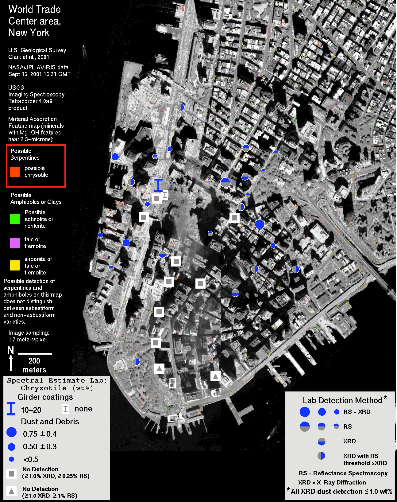
Mineralogic characterization of field samples (discussed in the laboratory sections) suggests that only trace levels of chrysotile asbestos were present in some of the samples from the WTC area, with one exception. The exception is a coating from a steel beam (sample WTC01-08) which showed chrysotile levels that could be as high as 20%. It should be noted that the field sampling sites do not coincide with AVIRIS pixel locations that indicate possible chrysotile or amphibole presence (A-Figure 4). But many field sample locations whose samples contain trace asbestos occur near locations in the AVIRIS map that indicate higher levels of serpentine minerals.
The correlation of mineralogically characterized field samples and AVIRIS mapped serpentine and amphibole mineralogy is shown in A-Figure 4. There appears to be a bias in chrysotile detection into a broad east-west trend, in both the AVIRIS and the laboratory results. This asymmetry in the dust/debris composition is supported by other compositional analyses, both AVIRIS and laboratory (see lab spectroscopy and integrations of results sections of this report).
The lower limit of detection of the AVIRIS instrument for asbestos minerals and, consequently, the WTC AVIRIS data set is probably in the few percent range as noted above. Asbestos levels below this detection limit and the implications of low levels of asbestos on human health are beyond the scope of this report.
The fact that field sampling shows high levels of chrysotile asbestos in some of the steel beam coatings (up to 20%) indicates the need for dust control during beam removal. Workers who are removing the beams should wear proper respiratory protection. However, video from news reports of the cleanup activity show that many of the steel beams no longer have the insulation coating. Where did the coatings go? It is probably now in the dust and debris. Thus the possibility exists that there may be other pockets of high levels of chrysotile, leading to the conclusion that workers should employ protective measures when cleaning up any dust or debris.
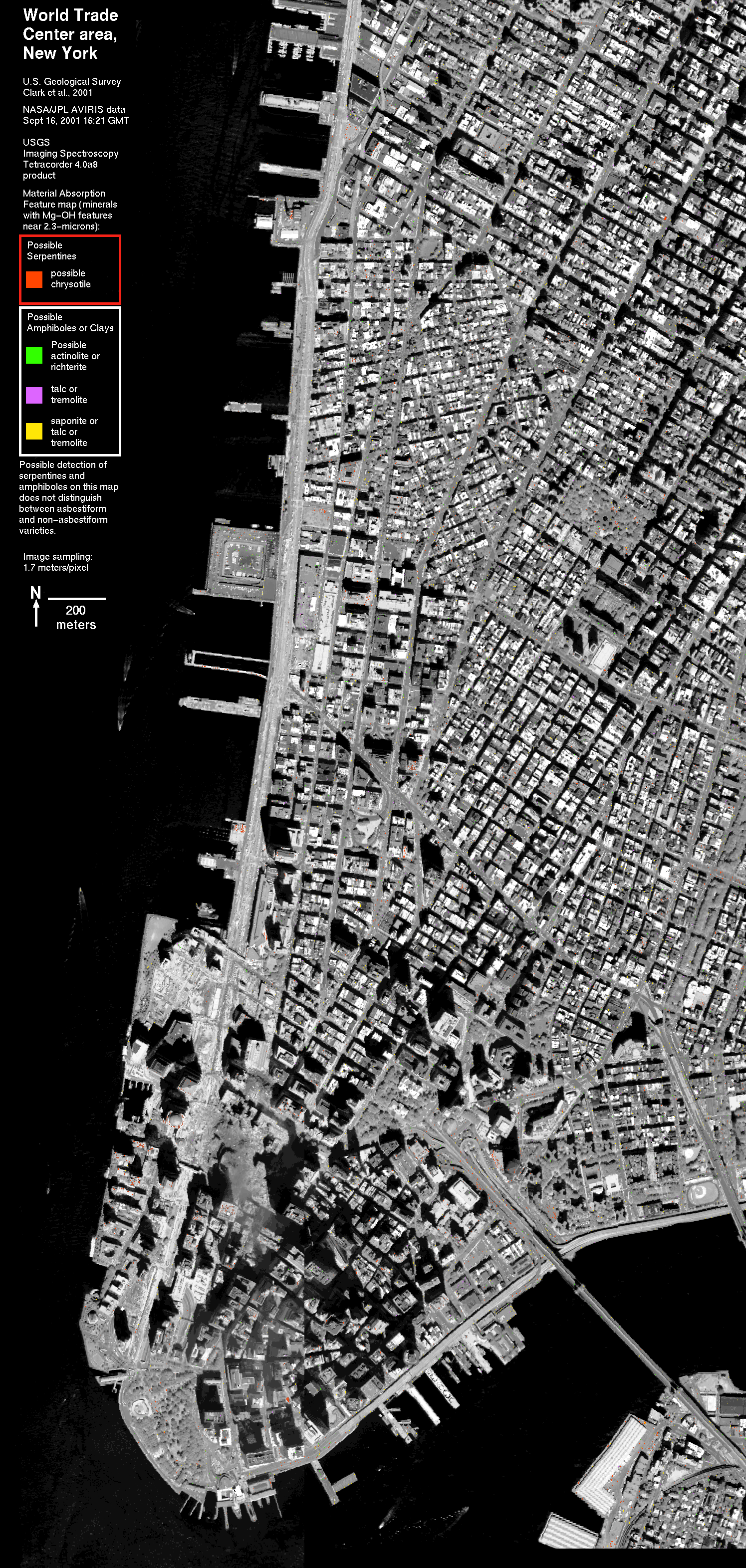 Larger 1751 KB image A-Figure 1a. Serpentine and Amphibole minerals map. |
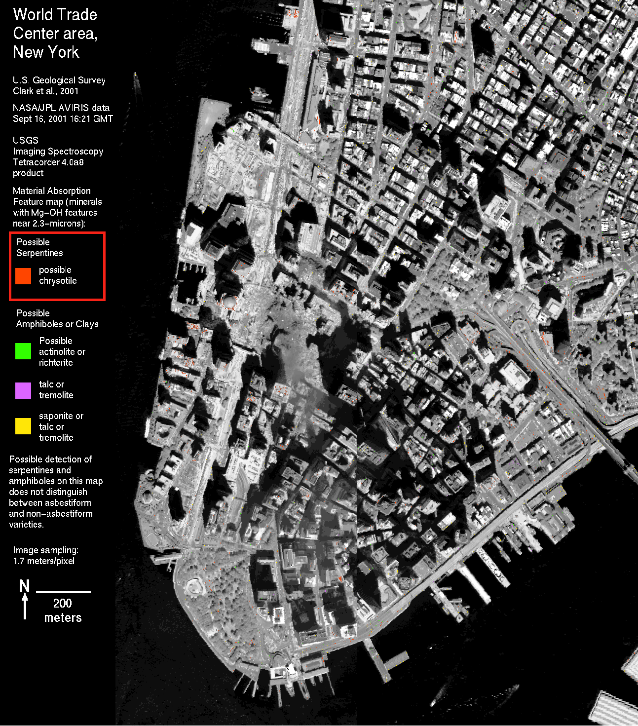 Larger 632 KB image A-Figure 1b. Serpentine and Amphibole minerals map, same as at right, but zoomed in to lower Manhattan. |
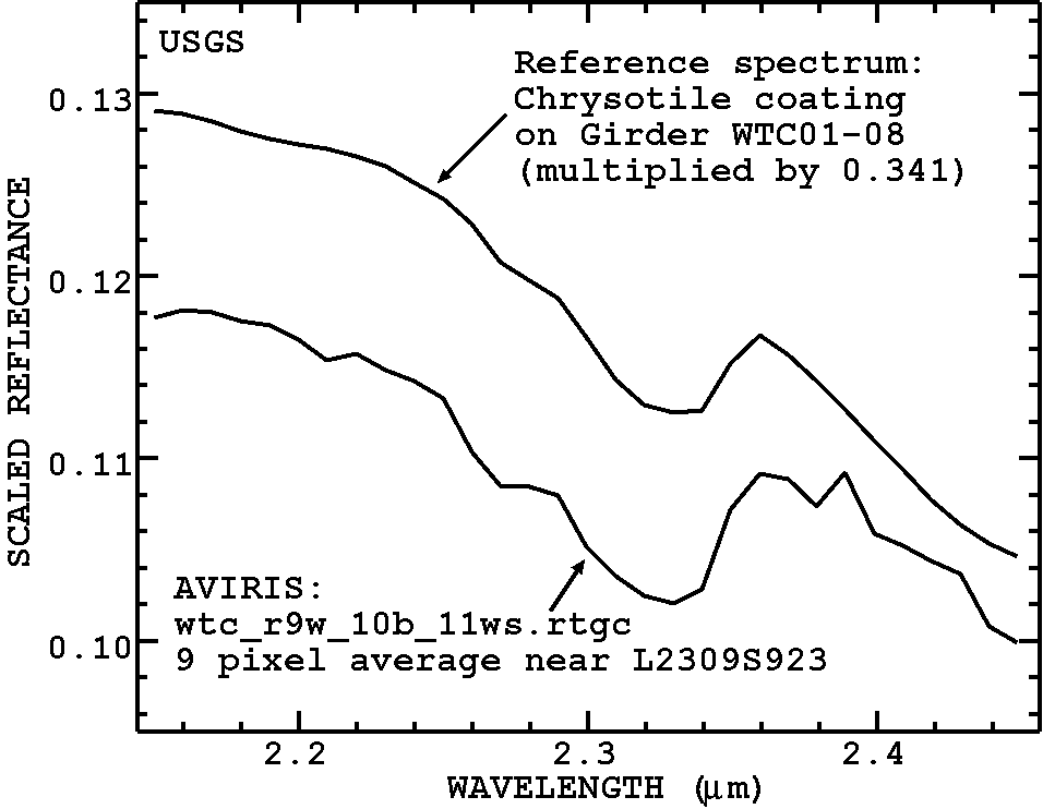
A-Figure 2. Spectra of some of the mapped chrysotile is a close match to the spectra of coating WTC01-08 which contains up to about 20% chrysotile.
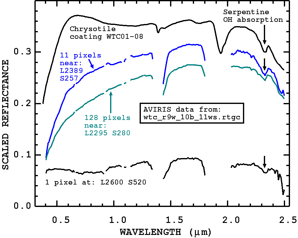
A-Figure 3. Example areas in the image with serpentine-like hydroxyl absorption feature.
A-Figure 4. WTC Sample analyses are shown plotted on the serpentine/amphibole AVIRIS map. There is a loose correlation of chrysotile locations spread in an east-west direction in both the laboratory analyses and the AVIRIS data.
NEXT PAGE of Report: Dust and Debris Plume Map
Back to document Table of Contents
For further information, contact:
Dr. Roger N. Clark
rclark@usgs.gov
| AccessibilityFOIAPrivacyPolicies and Notices | |
| |
|