FISC - St. Petersburg
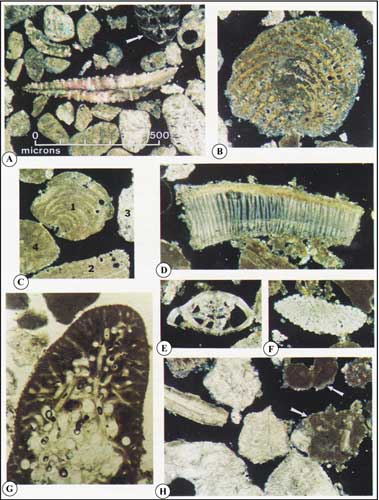
Figure 12. Petrographic-microscope photos show examples of thin-sectioned sand grains (from the Looe Key Reef area, Fig. 6A, Tile 6, lower Keys; from Lidz et al., 1985). Scale for all photos is the same; all photos except (E) are under cross-polarized light. (A) Center object is a spicule or stiffening skeletal element from a sea fan. Arrow points to the shell of a foraminifer, a calcifying protozoan. Most other grains are coral. (B) Foraminifer. (C) Grains 1 and 2 are red algae. Grain 3 is coral, 4 is Halimeda. (D) Molluscan fragment. (E) Foraminifer. (F) Sea-urchin fragment. (G) Section of whole Halimeda plate shows diagnostic internal tubes. (H) Coral grains. Arrows point to grains that have been partially or wholly altered to micrite (biologically altered to fine-grained carbonate).
|
Can't see the printable PDF version? Get the free Adobe Acrobat® Reader. |

![]() U.S. Department of the Interior |
U.S. Geological Survey
U.S. Department of the Interior |
U.S. Geological Survey
URL: [disc] /pubs/pp/2007/1751/professional-paper/figures/fig12.html
Page Contact Information: Feedback
Page Last Modified: December 01, 2016 @ 04:11 PM (JSS)
