 |
|
|
||||
| OFR 01-0429: World Trade Center USGS Integration of Results and Conclusions |
| About USGS / Science Topics / Maps, Products & Publications / Education / FAQ |
The results of analyses completed so far show a consistent picture: the samples are largely composed of gypsum, cellulose, and miscellaneous materials common in a building, with minor asbestiform minerals. However, one sample analyzed, the coating on a steel beam, indicates the presence of a significant abundance of chrysotile asbestos (as much as 20% by volume). The confirmed abundant chrysotile sample and the potential pockets of chrysotile indicated in the AVIRIS mineral maps indicates that asbestos can be found in localized concentrations. Thus, appropriate precautions should be taken when handling debris, especially coatings on metal beams.
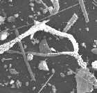
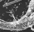
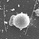
Sample results are summarized in Table 1, below. To see the full resolution SEM image and description, click on the image in the table. In the spectroscopy column in the table below, CH indicates organic compounds, including paints and plastics. Fe2+ indicates minerals or materials containing ferrous iron. Amounts are qualitative and indicate (from low to high) trace as (tr), weak as (wk), and strong as (str). No indication indicates between weak and strong.
The composition of samples collected in the WTC area, as indicated by spectroscopy, XRD, SEM, and from the visual examination during splitting of the samples, show similarities, yet each sample shows differences. Thus, while the samples appear to be a "grey dust", the data indicate the dust was not well mixed. The sample analyses and the AVIRIS mapping results agree in this regard.
| Sample Number | Spectroscopy | XRD | SEM | Leach pH |
Location |
| WTC01-01 (calibration) | asphalt from NJ | ||||
| WTC01-02 | gypsum, muscovite (tr), CH, Fe2+ |
Quartz - Minor Calcite - Minor Gypsum - Minor Dolomite - Trace Anhydrite - Trace Orthoclase - Trace *Major amorphous material |
10.1 | Water & Vietnam Vet. Plaza 4506052N 583559E |
|
| WTC01-03 | gypsum, muscovite (tr), CH, Fe2+ (wk) |
Quartz - Major Gypsum - Minor Calcite - Minor Anhydrite - Trace *Major amorphous material |
 |
9.51 | Battery Park, east central 4506123N 583217E drainage disc |
| WTC01-04 | gypsum, muscovite and/or portlandite (tr), CH, Fe2+ (wk) |
Calcite - Major Gypsum - Minor Anhydrite - Minor Quartz - Trace Muscovite - Trace Microcline - Trace *Major amorphous material |
Battery Park, NE end 4506284N 583233E |
||
| WTC01-05 | gypsum, muscovite and/or portlandite (tr), CH, Fe2+(wk) |
Calcite - Major Quartz - Minor Gypsum - Minor Anhydrite - Minor Calcium Sulfate Hydrate - Trace *Major amorphous material |
9.9 | Broadway & Wall St. from Bank of NY |
|
| WTC01-06 | gypsum, muscovite and/or portlandite (tr), CH, Fe2+ |
Calcite - Minor Quartz - Minor Gypsum - Minor Anhydrite - Minor Muscovite - Trace Calcium Sulfate Hydrate - Trace *Major amorphous material |
9.65 | Greenwich & Morris |
|
| WTC01-07 | gypsum, muscovite and/or portlandite (tr), CH, Fe2+ |
Gypsum - Minor Calcite - Minor Quartz - Minor Anhydrite - Minor Muscovite - Trace Microcline - Trace *Major amorphous material |
Rector & Greenwich from sidewalk |
||
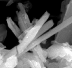
| WTC01-08 | chrysotile gypsum CH, Fe2+ (str) |
Calcite - Minor Chrysotile - Minor Gypsum - Minor Talc - Trace Anhydrite - Trace Microcline - Trace Rozenite - Trace *Possible trace rozenite *Major amorphous material |
 |
West End & Vesey, steel girder |
|
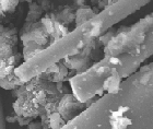
| WTC01-09 | gypsum, muscovite, CH, Fe2+ (str) |
Calcite - Minor Gypsum - Minor Thaumasite - Minor Quartz - Trace Calcium Silicate - Trace Larnite - Trace *Major amorphous material |
10.8 | West End & Vesey, steel girder |
|
| WTC01-10 | gypsum, muscovite (tr), CH, chrysotile (tr) Fe2+ |
Gypsum - Minor Calcite - Minor Quartz - Minor Anhydrite - Trace Muscovite - Trace *Major amorphous material |
West End & Vesey |
||
| WTC01-11 | gypsum, muscovite (tr), CH, Fe2+ |
Quartz - Minor Gypsum - Minor Calcite - Minor Anhydrite - Minor Microcline Trace Muscovite - Trace Chrysotile - Trace Calcium Sulfate - Trace *Major amorphous material |
West End & Barclay |
||
| WTC01-12 | gypsum, muscovite (tr), CH, Fe2+ |
Quartz - Minor Calcite - Minor Gypsum - Minor Anhydrite - Minor Muscovite - Trace Chrysotile - Trace *Major amorphous material |
West End & Warren |
||
| WTC01-13 | gypsum, muscovite (tr), CH, Fe2+ (wk) possible trace chrysotile |
Quartz - Minor Calcite - Minor Gypsum - Minor Anhydrite - Minor Muscovite - Trace *Major amorphous material |
Broadway & Barclay |
||
| WTC01-14 | gypsum, muscovite, CH, Fe2+ (wk), chrysotile (wk) |
Gypsum - Minor Calcite - Minor Quartz - Minor Anhydrite - Trace Muscovite - Trace *Major amorphous material |
9.68 | Church & Barclay from a windowsill |
|
| WTC01-15 | gypsum, muscovite, CH, Fe2+ (wk) possible trace chrysotile |
Gypsum - Major Anhydrite - Minor Quartz - Minor Calcite - Minor Muscovite - Trace *Major amorphous material |
10 | Maiden Ln. & Church |
|
| WTC01-16 | gypsum, muscovite and/or portlandite (tr), CH, Fe2+ (wk) |
Quartz - major Gypsum - Major Calcite - Minor Anhydrite - Trace Albite - Trace Orthoclase - Trace *Major amorphous material |
8.22 | Broadway & Dey |
|
| WTC01-17 | gypsum, muscovite and/or portlandite (tr), CH, Fe2+ |
Quartz - Minor Gypsum - Minor Calcite - Minor Anhydrite - Minor Calcium Sulfate Hydrate - Trace Muscovite - Trace *Major amorphous material |
9.47 | Greenwich & Albany |
|
| WTC01-18 | gypsum, portlandite (tr), CH, Fe2+ (wk) |
Gypsum - Minor Calcite - Minor Quartz - Minor Muscovite - Minor Anhydrite - Minor Portlandite - Trace Bassanite - Trace Chrysotile - Trace *Possible trace chrysotile *Major amorphous material |
Washington & Albany |
||
| WTC01-19 | gypsum, muscovite (tr), CH, Fe2+ (wk) |
Gypsum - Minor Calcite - Minor Quartz - Minor Muscovite - Trace Anhydrite - Trace Bassanite - Trace *Major amorphous material |
South End & Ceder |
||
| WTC01-20 | gypsum, muscovite and/or portlandite (tr), CH, chrysotile (tr), Fe2+ (wk) |
Quartz - Minor Calcite - Minor Gypsum - Minor Anhydrite - Minor Portlandite - Trace Bassanite - Trace Muscovite - Trace Clinochrysotile - Trace *Very slight possibility of a trace of chrysotile *Portlandite and bassanite are trace to minor *Major amorphous material |
 |
11.8 | Liberty & South End 2 World Financial Center: indoor sample |
| WTC01-21 | gypsum, muscovite, CH, chrysotile (tr) Fe2+ (wk) |
Gypsum - Minor Quartz - Minor Calcite - Minor Anhydrite - Minor Microcline - Trace Clinochrysotile - Trace Muscovite *possible trace Chrysotile *Major amorphous material |
9.98 | Directly West of WTC at ferry dock and ped. mall |
|
| WTC01-22 | gypsum, muscovite, CH, Fe2+ (wk) |
Gypsum - Major Anhydrite - Minor Quartz - Minor Calcite - Minor Muscovite - Trace Paragonite - Trace Lizardite - Trace Magnesionhornblende - Trace Clinochrysotile - Trace *possible trace (<.5%) chrysotile and/or Lizardite *trace amphibole (<.5%) *Major amorphous material |
 |
10.4 | 1/2 way between WTC and Ferry Dock along ped. mall from a windowsill |
| WTC01-23 | gypsum, muscovite (tr), CH, chrysotile (tr) Fe2+ (wk) |
Gypsum - Minor Muscovite - Minor Calcite - Minor Quartz - Minor Anhydrite - Trace Bassanite - Trace *Major amorphous material |
Broadway & Murray |
||
| WTC01-24 | gypsum, muscovite (tr), CH, chrysotile (tr) Fe2+ (wk) |
Gypsum - Minor Calcite - Minor Anhydrite - Minor Dolomite - Minor Muscovite - Minor Quartz - Trace Microcline - Trace Calcium Sulfate - Trace *Major amorphous material |
Broadway & Chambers |
||
| WTC01-25 | gypsum, muscovite/illite, CH, chrysotile (tr) Fe2+ (wk) |
Calcite - Minor Quartz - Minor Gypsum - Minor Anhydrite - Minor Dolomite - Trace Illite - Trace *Major amorphous material |
9.37 | Church & Warren |
|
| WTC01-26 | gypsum, muscovite (tr), CH, Fe2+ (wk) |
Calcite - Minor Gypsum - Minor Anhydrite - Minor Muscovite - Minor Quartz - Trace Dolomite - Trace Microcline - Trace Calcium Sulfate - Trace Chrysotile - Trace *Major amorphous material |
1/2 way between Broadway and Church on Park |
||
| WTC01-27 | gypsum, muscovite, CH |
Anhydrite - Minor Calcite - Minor Quartz - Minor Gypsum - Minor Orthoclase - Trace Albite - Trace Muscovite - Trace Clinochrysotile - Trace *Major amorphous material |
 |
10 | Broadway & Vesey |
| WTC01-28 | gypsum, muscovite, CH, chrysotile (wk), Fe2+ (wk) |
Calcite - Minor Gypsum - Minor Quartz - Minor Anhydrite - Minor Chrysotile - Trace *Major amorphous material |
9.93 | Ann & Nassau |
|
| WTC01-29 | gypsum, muscovite (tr), CH, chrysotile (tr), Fe2+ (wk) |
Calcite - Minor Gypsum - Minor Quartz - Minor Anhydrite - Minor Muscovite - Trace Chrysotile - Trace *Major amorphous material |
William & Fulton |
||
| WTC01-30 | gypsum, muscovite, CH, Fe2+ |
Calcite - Minor Quartz - Minor Dolomite - Minor Anhydrite - Minor Gypsum - Minor Chrysotile - Trace *Possible trace chrysotile *Major amorphous material |
Fulton & Cliff St. from on top of a car |
||
| WTC01-31 | gypsum, muscovite, CH, Fe2+ (wk) |
Calcite - Minor Gypsum - Minor Quartz - Minor Anhydrite - Minor Muscovite - Trace Chrysotile - Trace *Major amorphous material |
William & Platt |
||
| WTC01-32 | gypsum, muscovite, CH, Fe2+ |
Calcite - Minor Gypsum - Minor Quartz - Minor Anhydrite - Minor Muscovite - Trace Chrysotile - Trace *Possible trace chrysotile *Major amorphous material |
William & Pine |
||
| WTC01-33 | gypsum, muscovite (tr), CH, Fe2+ (wk) |
Gypsum - Minor Calcite - Minor Quartz - Minor Muscovite - Minor Anhydrite - Trace Magnesiohornblende - Trace Chrysotile - Trace *Major amorphous material |
Battery Park, middle |
||
| WTC01-34 | gypsum, muscovite, CH, chrysotile (tr) Fe2+ (wk) |
Quartz - Major Calcite - Minor Gypsum - Minor Anhydrite - Trace *Major amorphous material |
9.8 | South End & Thames |
|
| WTC01-35 | gypsum, CH, muscovite, chrysotile (tr) Fe2+ |
Gypsum - Minor Calcite - Minor Quartz - Minor Anhydrite - Trace Muscovite - Trace Chrysotile - Trace *Major amorphous material |
; | Albany & Hudson River |
|
| WTC01-36 | gypsum, muscovite and/or portlandite (tr), CH, Fe2+, possible trace chrysotile |
Gypsum - Minor Calcite - Minor Quartz - Minor Anhydrite - Minor Bassanite - Minor Portlandite - Minor Dolomite - Trace Illite - Trace Chrysotile - Trace *possible trace chrysotile *Major amorphous material |
 |
11.8 | South End & Albany (30th floor): Indoor sample |
| WTC01-37A | gypsum, muscovite, portlandite, Fe2+ |
Quartz - Major Albite - Major Portlandite - Minor Magnesiohornblende - Minor Orthoclase - Trace Muscovite - Trace |
concrete from WTC area | ||
| WTC01-37B | portlandite, Fe2+ |
Quartz - Major Orthoclase - Minor Portlandite - Minor Albite - Trace Calcite - Trace Magnesiohornblende - Trace Muscovite |
concrete from WTC area |
* Amorphous material is not identifiable by XRD, but its presence is detectable.
* "Possible trace chrysotile" means at or near the detection limit with XRD.
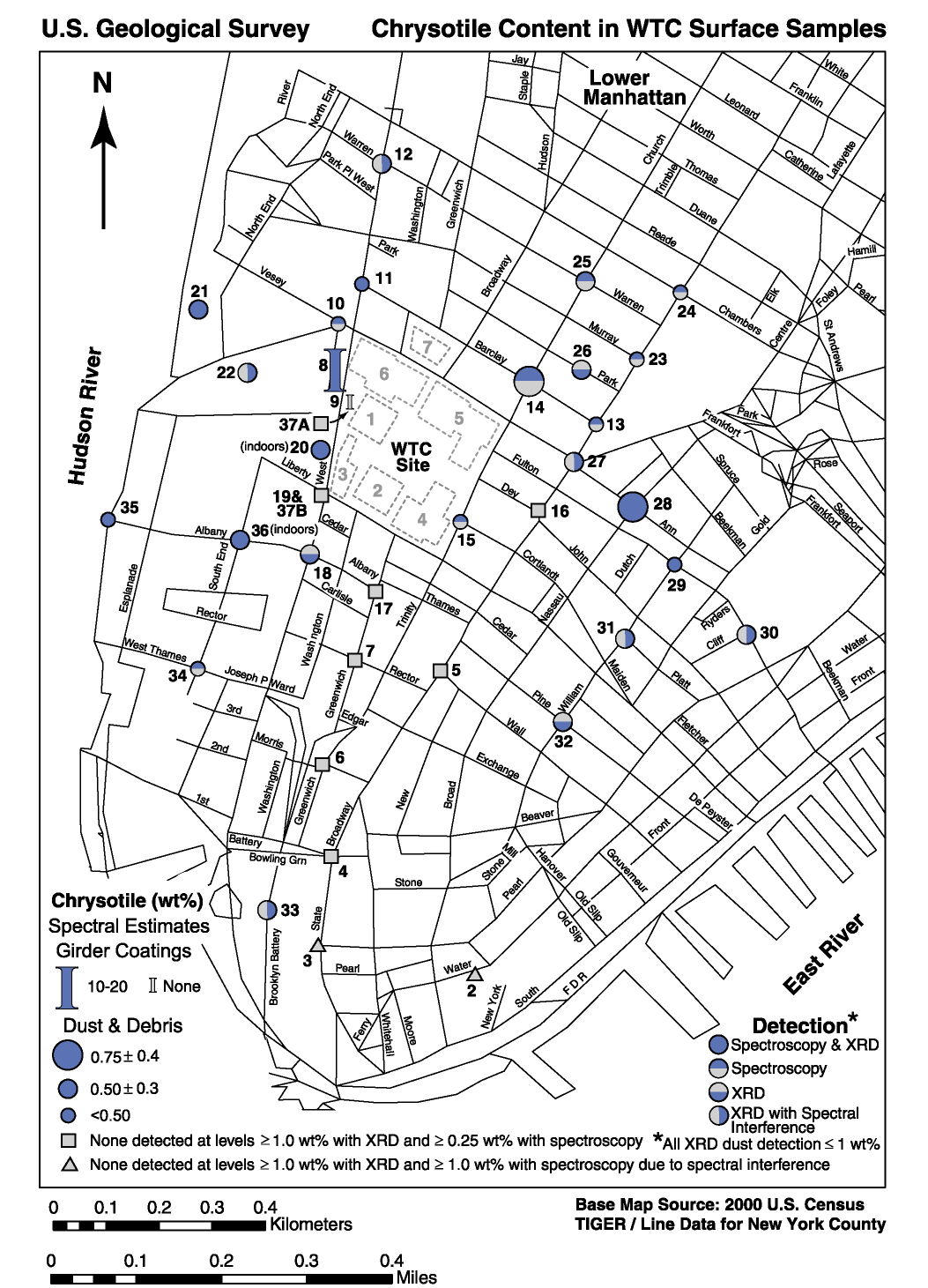
The question of asbestos distribution was investigated and the results show an asymmetric distribution pattern (Results Figure 1). More chrysotile was detected in an east-west direction than south. This pattern occurs in both the AVIRIS maps and from field samples (Results Figure 2). While there is a general trend, it is not exclusive, meaning that chrysotile was detected in all directions. It also should be noted that samples obtained next to each other (on the map this means a city block apart) can show different results: one has asbestos, another has no chrysotile above the detection limit.
Composition of samples on a centimeter scale was examined with a spectrometer. Small variations in chrysotile content throughout a sample were observed. Thus from scales of cm to tens of meters, chrysotile content varies. Such variability makes sampling and overall assessment of a site difficult.
The fact that some materials in the WTC debris were observed to contain higher levels of chrysotile (sample WTC01-08) on a steel beam, and that the coatings on the beams have largely been stripped, leads to the question of where did the coatings go and how well distributed/dispersed is the chrysotile? Because a patch of coating showed up to 20% chrysotile, and the field samples and the AVIRIS maps show varying levels of serpentine (chrysotile) leads to the possibility that other patches of chrysotile may exist in the debris.
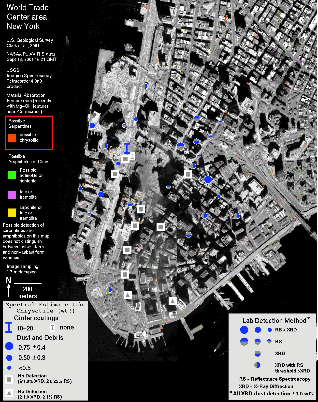
The asymmetry in the AVIRIS iron-bearing materials map may be related to the asymmetry in the asbestiform minerals map. The AVIRIS data and the laboratory analyses of the field samples indicate a lower abundance of chrysotile in the the southern direction from the WTC, the same direction of the increase in iron-bearing materials. The one field sample, WTC01-08, from an iron beam, which had up to 20% chrysotile also contains a strong Fe2+ absorption. Thus one might expect a higher chrysotile content in iron-bearing materials. However, this is clearly not the case, at least in general. This may indicate other sources of the chrysotile besides the beam coatings.
AVIRIS imaging spectroscopy mapping provides a synoptic view that samples more area than possible with other methods. The AVIRIS maps shown here represent only a portion of the data collected, and effectively provide data for about 4.7 million sample locations, all obtained within a couple of hours. The sampling includes land, air and water.
The fact that the field sampling missed the highest concentrations of serpentines in the AVIRIS maps shows the limitations of limited sampling methodologies. Ideally, the field sampling team would have the AVIRIS materials maps to guide the field sampling. Unfortunately, this was not possible in this rapid response case (but we routinely employ such methods in geologic studies where the region does not change rapidly). Even so, the materials maps for this study were produced faster than any other imaging spectroscopy effort to our knowledge. The AVIRIS data were received within 24 hours of acquisition, and the data were initially calibrated to help the field team obtain the final calibration data with real time feedback via cell phone. In this case, scientists in Denver communicated composition of field calibration sites using initially calibrated AVIRIS data (of the parking lot structure) while the field team was investigating where the best portion of the parking lot was located. The real-time feedback resulted in avoidance of portions of the parking lot with strong absorption features, not visible to the human eye, that could have compromised the quality of the final calibration.
With further development of on-board solar calibration targets on the aircraft with the AVIRIS sensor, the refinement of analysis software, the development of more reference spectral libraries, and the use of faster computers, an even faster response is possible in the future. The challenge is formidable. To analyze the data for this study, we used approximately 300 gigabytes of disk space and performed over 50 trillion calculations. The results of the AVIRIS mapping are limited by knowledge of the spectral properties of materials and the detection levels are limited by the sensor signal-to-noise. The detection limits could be substantially improved with existing technology in a new sensor design.
The combination of field sampling with laboratory analysis and imaging spectroscopy remote sensing provide a powerful assessment combination. We estimate the analysis effort of this highly experienced team to be 1.8 person years to complete this study plus another 0.6 person-year for the AVIRIS data collection effort. This study includes analysis of 20% of the AVIRIS data from Sept 16, and 7% of the data from Sept 23 (thermal hot spot analysis only).
The scientific data from this study is presented with no assessment of health effects. It is beyond the scope of this study to assess health effects of a fraction of a percent chrysotile asbestos, for example.
Other Conclusions have already been presented in the Executive Summary.
Results Figure 1. Sample location map coded by asbestos detection.
Results Figure 2.WTC Sample analyses from Results Figure 1 are shown plotted on the serpentine/amphibole AVIRIS map. There is a loose correlation of chrysotile locations spread in an east-west direction in both the laboratory analyses and the AVIRIS data.
NEXT Section of Report: References
Back to document Table of Contents
| AccessibilityFOIAPrivacyPolicies and Notices | |
| |
|