Biogeochemical Processes That Produce Dissolved Organic Matter From Wheat StrawBy Robert L. Wershaw, David W. Rutherford, Jerry A. Leenheer, Kay R. Kennedy, Larry G. Cox, and Donald R. Koci |
|||||||||||||||||||||||||||||||||||||||||||||||||||||||||||||||||||||||||||||||||||||||||||||||||||||||||||||||||||||||||||||||||||||||||||||||||||||||||||||||||||||||||||||||||||||||||||||||||||||||||||||||||||||||||||||||||||||||||||||||||||||||||||||||||||||||||||||||||||||||||||||||||||||||||||||||||||||||||||||||||||||||||||||||||||||||||||||||||||||||||||||||||||||||||||||||||||||||||||||||||||||||||||||||||||||||||||||||||||||||||||||||||||||||||||||||||||||||||||||||||||||||||||||||||||||||||||||||||||||||||||||||||||||||||||||||||||||||||||||||||||||||||||||||||||||||||||||||||||||||||||||||||||||||
MATERIALS AND METHODSSample collectionThe black water from decomposing wheat straw was collected in Teflon bottles from pools in a field near Hutchinson, Kansas, where bales of wheat straw were stacked. The bottles were packed in ice and shipped overnight to a USGS laboratory in Denver, Colo., where they were immediately refrigerated. Wheat straw was purchased from a local feed dealer in the Denver area. Dissolved organic carbon and inorganic analysesDissolved organic carbon (DOC) measurements were made by persulfate wet oxidation in a gas-tight reaction vessel in an Oceanography International (OI) Model 700 carbon analyzer (Aiken, 1992). Samples were introduced into the reaction vessel through a fixed-volume sample loop. Prior to oxidation, the samples were acidified with phosphoric acid and purged with nitrogen to remove dissolved carbon dioxide. Inorganic cation concentrations in the black water were measured by inductively coupled plasma-emission spectrometry. The black water was filtered through a 0.45-micrometer filter prior to analysis. Carbohydrate analysisThe composition of the carbohydrate polymers in the black water DOM was determined by gas chromatographic analysis of the alditol acetates of the acid hydrolyzate of the DOM by V-Labs (Covington, Louisiana 70433) using the method described by Albersheim and others (1967). A dried, weighed sample was hydrolyzed in dilute trifluoroacetic acid followed by reduction with sodium borohydride and acetylation with acetic anhydride. The resulting alditol acetates were separated by gas-liquid chromatography and quantitated by comparison with calibration standards. Free and bound carbohydrates in a sample were measured colorimetrically by the phenol-sulfuric acid test (Dubois and others, 1956). A weighed sample was first shaken vigorously with water, and an aliquot of the supernatant was reacted with the phenol-sulfuric acid reagent and analyzed. This yielded a measure of free carbohydrate in the sample. The bound carbohydrates were measured by reacting the suspension with the phenol-sulfuric acid mixture. FractionationThe DOM in the water was fractionated into four fractions of increasing polarity using a modification of the procedure described by Wershaw and others (1996). In this procedure the black water without pH adjustment was first pumped into a glass column with fluorocarbon fittings filled with 150 milliliters (mL) of XAD-8 resin, and the column was then rinsed with 300 mL of deionized water. The adsorbed material (fraction 1) was eluted with 100 mL of a solution of 75 percent by volume acetonitrile in water. The eluate was evaporated to dryness in a rotary evaporator, the DOC was dissolved in water and then lyophilized. The combined black water and rinse water that passed through the column in the first step of the procedure was acidified to pH 2 with hydrochloric acid (HCl) and pumped through the column again followed by 300 mL of 0.01 molar (M) HCl. The fraction adsorbed (fraction 2) was eluted and lyophilized as above. The pH of the combined black water and rinse solution that passed through the column in the second step was adjusted to 5 with NaOH, and the volume was reduced to 500 mL by rotary evaporation. The pH was then lowered to 2, and the concentrated solution was again pumped onto the XAD-8 column. The fraction adsorbed (fraction 3) was eluted and lyophilized. The pH of the solution of residual DOC that did not adsorb on the XAD 8 was adjusted to 2 with 1 M HCl and pumped through a 100 mL XAD-4 column. The column was eluted with 100 mL of 75 percent by volume acetonitrile in water, and the eluate was concentrated to 20 mL by rotary evaporation. The residual HCl and water were removed by repeated additions of anhydrous acetonitrile followed by evaporation. The dried sample (fraction 4) was dissolved in 50 mL of water and lyophilized. In all of the operations above a positive displacement pump with a stainless steel body, ceramic piston, and sapphire ball valves and fluorocarbon tubing were used. Therefore, the solutions only came in contact with glass, fluorocarbon, stainless steel, ceramic, and sapphire. The relative concentrations of the fractions obtained in one of the fractionation runs were: fraction 1, 31 percent; fraction 2, 53 percent; fraction 3, 14 percent; and fraction 4, 2 percent. The humic acid fraction (designated here as black water precipitate or BWP) was isolated by adjusting the pH of a 30 mL aliquot of the black water to 1 with 1 M HCl and then centrifuging the resulting suspension. The supernatant was poured off and the precipitate was dispersed in 20 mL of water and re-centrifuged. The supernatant was discarded, and the precipitate was washed and centrifuged two more times; the supernatant was discarded after each washing. The pH of the supernatant after the last washing was between 3 and 4. The precipitate was re-suspended in water and lyophilized. This procedure yielded 0.1406 gram (g) of humic acid (84 percent of the DOC in the water). DialysisExhaustive dialysis of the black water was carried out by placing approximately 50 mL of the black water in a dialysis bag made of SpectraPor® 3 regenerated cellulose dialysis casing (Spectrum Laboratories, Rancho Dominguez, Cal.). Two different casings were used; one had a nominal 3,500 dalton (Da) molecular-weight cutoff and the other a cut off of 12,000 Da (calibrated with globular proteins); both casings were 29 millimeters (mm) in diameter. Each dialysis bag was suspended in a 1 liter (L) graduated cylinder filled with de-ionized water. The water in the cylinder was constantly stirred with a magnetic stirrer, and changed daily until no more color diffused out of the bag. Spectral analysesSolid-state 13C NMR spectra of the lyophilized fractions were measured on a Chemagnetics CMX 200-megahertz (MHz) proton frequency spectrometer (Wershaw and others, 1998) equipped with a 7.5 mm Chemagnetics ceramic probe. The samples were packed in zirconia rotors and spun at 5000 hertz (Hz). The acquisition parameters were contact time of l millisecond (ms) or 5 ms, pulse delay of 1 second (s), and 4.5 microsecond (µs) 90º pulse. Liquid-state 13C NMR spectra of the humic acid fraction dissolved in d6 methyl sulfoxide (Aldrich Chemical Co., 99.5 atom percent deuterium) were measured on a 500 MHz JEOL Eclipse spectrometer. The refocused insensitive nuclei enhanced by polarization transfer (INEPT) pulse sequence was used. The proton frequency was 500 MHz and the carbon frequency was 125 MHz. The measurements were made at constant temperature of 50º Centigrade (C); the following parameters were used: WALTZ proton decoupling with an offset of 5.0 parts per million (ppm), J constant of 140 Hz, carbon acquisition time of 0.521 s, 13C offset of 100 ppm. A total of 16,384 data points were collected; the data were plotted with a line-broadening of 100 Hz. Infrared (IR) spectra of the fractions were measured on a Perkin-Elmer 2000 Fourier transform spectrometer. Pellets for IR analysis were prepared by triturating with a mortar and pestle approximately 5 milligrams (mg) of sample with about 250 mg of potassium bromide (KBr), and then pressing the mixture in a die. The NMR and IR spectra of the wheat straw were measured using the same methods as those used for the DOM fractions. Ultraviolet-visible (UV-visible) spectra were obtained with a Spectral Instruments (Tucson, Ariz.) 400 Series spectrophotometer fitted with a 1 centimeter (cm) path length fiberoptic dip cell. Molecular weight measurementsHigh-performance size-exclusion chromatography (HPSEC) with multi-angle laser-light scattering (MALLS) detection was performed at Chromaceutical Advanced Technologies, Inc., Hopkinton, Mass. A Waters Alliance 2695 HPSEC system with a Waters 2996 photodiode array detector, Wyatt Optilab DSP refractive index detector, and Wyatt MiniDawn light scattering detector was used. The column was a Tosoh Biosep GMPWx1 column 7.8 mm in diameter and 300 mm in length; the mobile phase was phosphate-buffered saline solution at pH 7.4 (0.138 molar sodium chloride, 10 millimolar phosphate, 200 ppm sodium azide). The flow rate was 1 mL/min, and the injection volume was 100 µL. The column temperature was maintained at 35ºC, the detector temperature at 40ºC, and the sample temperature at 25ºC. Two samples were subjected to HPSEC/MALLS analysis: untreated black water and the retentate from the exhaustive dialysis through the 12,000 Da membrane. The retentate was lyophilyzed after dialysis and then reconstituted in the phosphate-buffered saline solution for analysis. From the MALLS data it is possible to calculate the number-average molecular weight (Mn), weight-average molecular weight (Mw), the z-average molecular weight (Mz), and the root-mean-square (RMS) radius of the particles in solution (Wyatt, 1993). The ratio Mw/Mn provides a measure of the polydispersity of the particles in solution. BIOCHEMICAL STUDY RESULTSElemental compositionAnalysis of the black water (BW) yielded a DOC of 2,900 mg/L. This DOC is very much higher than one normally finds in a natural water. The pH of the BW measured in the laboratory was 7.7. The elemental composition of the BW is given in Table 1.
Lowering the pH of the black water to pH 1 yielded a precipitate (humic acid) that contained 84 percent of the DOC in the black water. The elemental composition of the black-water precipitate (BWP) reported here is considerably different from that of alkali lignin (AL) or milled wood lignin (MWL) isolated from wheat straw (Table 2). The higher oxygen content and lower carbon content of the BWP compared to the wheat-straw AL and MWL indicate that the BWP contains significant carbohydrate material, as will be demonstrated in the following section. The higher nitrogen content of the BWP probably represents amino acids and proteins derived from the wheat straw. The concentrations of carbohydrates, proteins, and amino acids are apparently reduced during the lignin isolation procedures.
Results of carbohydrate analysisCarbohydrate analyses of the BWP were performed. Phenol-sulfuric acid analysis of the water extract of BWP indicated that there are no free carbohydrates. When the 18 M sulfuric acid was added to the BWP suspension all of the suspended material went into solution. Colorimetric analysis indicated that 27 percent of BWP is insoluble carbohydrate. The results of specific carbohydrate analysis after hydrolysis with trifluoroacetic acid and gas-liquid chromatographic analysis of the alditol acetates are as follows: rhamnose 6 percent, arabinose 34 percent, xylose 32 percent, mannose 8 percent, galactose 7 percent and glucose 14 percent. These results indicate that the major carbohydrate polymer present is an arabino-xylan, and indeed, Lawther and others (1995) pointed out that wheat straw hemicellulose is composed mainly of b-1-4 linked D-xylopyranose units with side chains composed of L-arabinose, D-glucuronic acid, D-glucuronic-4-O-methyl ether, D-galactose, and possibly D-glucose units. Calibration of the gas-liquid chromatographic results with standards indicated that the total carbohydrate content of the BWP sample is 35 percent. This result is probably more accurate than the 27 percent result obtained colorimetrically. Ultraviolet-visible, nuclear magnetic resonance and infrared spectral resultsThe UV-visible spectrum of the BW (figure 2) has a well-defined absorption band at 278 nanometers (nm) which has been shown to be characteristic of lignin (Sun and Tomkinson, 2002; Scalbert and others, 1986). Studies with model compounds show that a band in this region is indicative of nonconjugated phenolic groups (Goldschmid, 1971). Sun and Tomkinson (2002) and Scalbert and others (1986) also found a band near 320 nm in the spectra of alkali lignins isolated from wheat straw. They attributed this band to hydroxycinnamic acids conjugated to phenolic groups. Only a weak hump in this region is present in the spectrum of the BW indicating that these conjugated structures are in lower concentrations in the BW than in the wheat-straw alkali lignins. The reduced intensity of this band probably indicates that some of the lignin groups are attached to non-phenolic groups such as carbohydrates (see Sun and Tomkinson, 2002).
Figure 2. Ultraviolet-visible spectrum of black water. The NMR spectra of the BWP, the four major fractions of the wheat straw DOM, and of dry, undegraded wheat straw are shown in figure 3. A generalized list of the types of functional groups that are represented by the various regions of DOM spectra is given in Table 3.
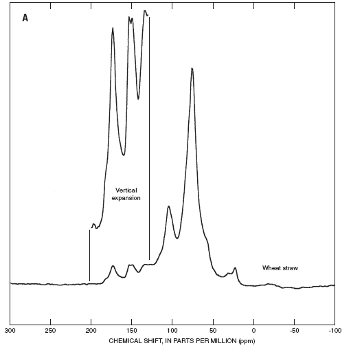
Figure 3. (A) 13C NMR spectrum of wheat straw (one millisecond contact time), and (B) 13C NMR spectra of black-water fractions (one millisecond contact time).
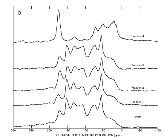
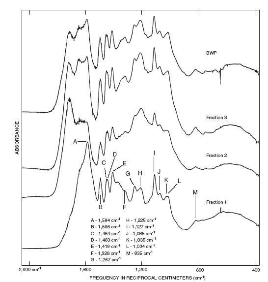
Figure 3. Continued. The most prominent aromatic carbon bandsin the wheat straw spectrum are at 153, 148, and 134 ppm. Hassi and others (1987) have shown that these bands are characteristic of guaiacylpropanoid and syringylpropanoid lignin monomeric units. The 153 ppm band represents the C-3 and C-5 carbon atoms of syringylpropanoid units in which there is an ether linkage at C-4. The 148 ppm band represents either the C-3 and C-4 carbons of guaiacylpropanoid units or the C-3 and C5 carbons of syringylpropanoid units in which C-4 is a phenol. The shoulder at 56 ppm probably represents methyl ether groups in lignin units. Evidence of lignin structures is most apparent in the spectra of fractions 1, 2, 3, and BWP where the aromatic bands are much more prominent than they are in fraction 4. The 152 ppm band is the strongest aromatic band in these spectra. A shoulder at about 148 ppm is present in the spectra of fractions 2 and 3. Broad bands between about 129 and 134 ppm also are present in the spectra of the first three fractions. Each of the spectra also has a broad band centered at about 108 ppm. The 130-134 ppm bands probably represent the C-1 and C-4 carbons of syringylpropanoid units. These bands occur between 136-138 ppm if C-4 is an ether. However, if C-4 is a phenol then C-1 has a chemical shift of 132 ppm and C-4 a shift of 135 ppm, this might lead one to conclude that C-4 linkages in the fractions and BWP are phenolic. Unfortunately, however, the strong 152 ppm band implies that C-4 is an ether linkage rather than a phenolic linkage. At the present time, this seeming contradiction cannot be explained. The bands centered at about 108 ppm most likely arise from C-2 and C-6 carbons of syringylpropanoid units in which C-a is attached to an oxygen atom (Scalbert and others, 1986). The INEPT data discussed below also indicate that C-a is attached to an oxygen atom. The bands at about 73 ppm in the spectra of fractions 1, 2, 3 and BWP probably represent carbohydrate units conjugated to the lignin as discussed below. The 56 ppm band, which is the strongest band in each of the spectra, indicates that the lignin has not undergone demethylation. Comparison of the ratios of the intensities of the 152 ppm bands to the intensities of the 148 ppm bands (or shoulders) in the spectra of fractions 1, 2, and 3 and BWP to the 152 ppm:148 ppm ratio in the undegraded wheat straw spectrum indicates that syringyl units are in greater concentration than guaiacyl units in the lignin of the BW isolates than in the wheat-straw lignin. This is consistent with the findings of Almendros and others (1992) that the residual lignin in decaying wheat straw is enriched in guaiacyl units. They pointed out that syringyl lignin components are generally less condensed and less resistant to degradation than guaiacyl components. The syringyl components, therefore, will be more readily leached from the decomposing wheat straw than the guaiacyl components. Carbonyl bands between 172 and 175 ppm are present in the NMR spectra of the wheat straw, the precipitate, and all of the fractions. These bands are relatively broad, and probably represent mixtures of aliphatic acids and esters. Fraction 1 was isolated at the ambient pH of the black water (pH 7.7), and therefore it must also contain some ionized carboxylate groups. More definitive results are provided by the IR spectra. The IR spectra of the samples provide further evidence of the chemical structures of the samples (figure 4). The IR spectrum (not shown) of the undegraded wheat straw has a band at 1,732 reciprocal centimeters (cm-1) that is consistent with the presence of aliphatic esters. A distinct carbonyl band is absent from the IR spectrum of fraction 1, however, a broad shoulder centered near 1,650 cm-1 is present. This shoulder and the band at 1,594 cm-1 probably represent the asymmetric stretching modes of different carboxylate groups. Fractions 2 and 3 and BWP have well-resolved carbonyl bands at about 1,720 cm-1 that are most likely representative of aliphatic carboxylic acids. Lignin extracts of wheat straw generally have fewer carboxylic acids groups than the BW isolates. For example, the carbonyl band in the spectrum of the acid-insoluble fraction of the potassium-hydroxide extract of wheat straw (wheat-straw alkali lignin) measured by Sun and Tomkinson, (2002) is only present as a weak shoulder at 1,702 cm-1. Well-resolved bands characteristic of lignins (Faix, 1991) are in the fingerprint regions of the IR spectra of BWP and of fractions 1-3. The 1,127 cm-1 band indicates that the lignin contains both guaiacyl and syringyl units, and the shoulder at about 1,160 cm-1 is indicative of p-hydroxyphenyl units; this shoulder was observed by Sun and Tomkinson (2002) in the IR spectrum of wheat-straw alkali lignin. The bands between 1,070 and 1,030 cm-1 in the BWP and fractions 1-3 spectra most likely represent carbohydrates (Colthup and others, 1990). The relative intensities of these two bands are similar to those of the carbohydrate bands in wood.
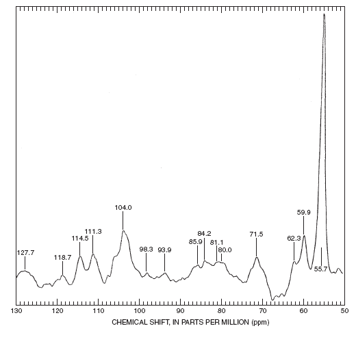
Figure 4. Infrared spectra of black water (BW) fractions 1. 2, 3 and black water precipitate (BWP). The liquid-state refocused INEPT spectrum of BWP, which only shows protonated carbon atoms, provides further support for the presence of lignin and carbohydrate units (figure 5). The acquisition parameters of the INEPT experiment have been chosen such that all of the protonated carbon atoms will be seen in the spectrum; however, the response is not quantitative. All of the bands in the spectrum can be assigned to hydroxyphenylpropanoid lignin or carbohydrate units, as shown in table 4. The bands at 59.9, 71.5, and between 80-86 ppm are all indicative of linkages between the b-carbon atoms of the propanoid side chains and the C-4 carbons of the aromatic rings (b-O-4) in the lignin units. No other types of linkages are detected in the INEPT spectrum. These results are consistent with the work of Terashima and others (1997) who found that between 65 and 74 percent of the linkages in wheat straw lignin are b-O-4, and that other types of linkages are in much lower concentrations. The sensitivity of the INEPT experiment in figure 5 apparently was inadequate to observe the other types of linkages. The 62.3 ppm band has a chemical shift very close to that attributed to the C-5 of xylose in hemicelluloses of barley straw lignin isolates (62.8 ppm) by Sun and others (2001).
Biogeochemical process implications of the spectral dataThe IR and NMR spectra of all of the BW isolates except fraction 4, which comprises about 2 percent of the NOM in the BW, have spectral bands characteristic of lignin. The NMR spectrum (figure 4) and IR spectrum (not shown) of fraction 4 indicate that it is most likely composed of a mixture of amino acids, sugar acids, and other aliphatic acids. This fraction probably represents a more complete degradation of the chemical components of wheat straw than the other fractions (Wershaw and others, 1996). The carboxylic acid bands in the NMR and IR spectra of the BW isolates probably represent more than one type of carboxylate group. Lam and others (1992) have shown that cinnamic acids linked to carbohydrate and lignin groups are present in wheat straw. Additional carboxylate groups may arise from the microbial degradation of lignin. The microbial degradation of wheat straw lignin has been found to involve oxidative depolymerization, preferential removal of syringyl groups, and an increase in carbonyl and carboxylate groups (Almendros and others, 1992; Crestini and others, 1998; Chen and Chang, 1985; Gaines and others, 1996; Vane and others, 2001). Crestini and others (1998) have proposed that wheat-straw lignin also may undergo aromatic-ring cleavage during the early stages of biodegradation. Thus, the aliphatic carboxylic acid bands in fractions 2-4 and the BWP probably represent both cinnamic acids and carboxylates formed during aromatic-ring cleavage. Aromatic-ring cleavage proceeds by the b–ketoadipate pathway. In this reaction sequence, ring cleavage takes place between two adjacent (ortho) hydroxyl groups on an aromatic ring which are oxidized to carboxylates. The resulting muconic acid (or muconate) undergoes isomerization to muconolactone followed by hydrolysis and further oxidation to b–ketoadipate. All of the steps in this sequence are enzymatically mediated as described in detail by Gaines and others (1996). Further oxidation (b-oxidation) produces succinyl-CoA and acetyl-CoA. Molecular weight of dissolved organic matterThe total organic carbon content (TOC) of the black water is 2.9 g/L which corresponds to about 5.8 g/L DOM assuming about 50 percent C in the DOM (see Table 2). This high concentration of the lignin-derived organic matter (OM) in the black water would initially appear to indicate this OM is made up of low molecular weight compounds. However, exhaustive dialysis through a 3,500 and 12,000 Da cutoff membrane indicated that 90 percent of the TOC would not penetrate the 3,500 Da membrane, and that 68 percent would not penetrate a 12,000 Da membrane. Results of HPSEC/MALLS analysis of the DOM in the black water and 12,000 Da retentate are listed in table 5.In principle, the MALLS technique, which measures scattered-light intensity as a function of angle, provides an absolute measure of the sizes of the particles in solution. However, the calculations used to obtain the values listed in the table require the measurement of other optical parameters such as the refractive index increment in order to yield accurate results (see Wyatt, 1993, for a detailed discussion). The results in Table 5 indicate that both samples are polydisperse and composed mainly of relatively high molecular weight particles. The dialysis has reduced the polydispersity of the DOM; however, in both samples there is a substantial amount of very high molecular weight particles as indicated by the very high Mz values. The relatively low RMS radius compared to Mn and Mw values of the black-water DOM strongly suggest that the particles in solution are mainly compact spheroidal structures. The increase in size of the dialyzed sample probably indicates that the constituent molecules of the lyophilized material did not fold into their most compact configurations when re-dissolved. The lower Mn , Mw, and Mz values of the 12,000 Da retentate compared to those of the black water probably are the result of differential solution with the highest molecular weight molecules not re-dissolving completely after freeze drying. Any undissolved material would be removed by the filtration prior to the chromatography.
Molecular weights from 324 to 17 x 106 Da have been reported for lignins extracted from wood (Goring, 1971). The results tabulated by Goring (1971) indicate that polydispersities (Mw/Mn) as high as seven have been observed. There are at least two possible explanations for these results. One possibility is that the molecular weight measurements accurately reflect the actual molecular weight distribution of the lignin polymer in wood. The other possibility is that the lignin is present in the wood as a high-molecular weight polymer, and that the relatively harsh extraction procedures used to isolate lignin from woody tissue partially degrade the polymer, yielding fragments of widely varying size. More recently, Goring (1989) has proposed that most lignin molecules are relatively low-molecular weight, two-dimensional networks that can form molecular aggregates. Molecular weight measurements by Pan and Sano (2000) and Evtuguin and others (2001) provide support for this hypothesis. They found that the molecular weights (either Mw or Mn) of lignin preparations isolated by such commonly used procedures as milling, alkaline, dioxane, or acetic acid extraction generally do not exceed 5,000 Da. Pan and Sano (2000) measured the molecular weights of lignin isolated from wheat straw by three different methods—milling, acetic acid extraction, and alkaline extraction at 160ºC by size-exclusion chromatography (SEC). The isolates were acetylated prior to the chromatography; polystyrene molecular-weight standards were used. The Mw values of the acetylated lignin isolates ranged from 3,270 to 4,430 Da and the Mn values from 1,770 to 2,020 Da. Sun and Tompkinson (2002) measured similar molecular weights by SEC on alkaline-extracted wheat straw lignin. Consideration of the way lignin forms in woody tissue provides a possible explanation for the large discrepancy between the measurements in this report and those of Pan and Sano (2000) and Sun and Tompkinson (2002). In woody terrestrial plants lignin is imbedded in the hemicellulose matrix of the cell walls. Lignin deposition is one of the final stages of xylem-cell differentiation (Donaldson, 2001). The deposition takes place in the interlamellar voids of the cell walls. During the deposition, chemical bonds form between the lignin and hemicellulose carbohydrates (Sun and others, 2000). Lam and others (1992) have shown that cinnamic acid groups in wheat lignin are bound to polysaccharides by ester linkages. Two different mechanisms have been proposed for the lignin polymerization. Lewis and his co-workers (see Burlat and others, 2001) have proposed that lignin deposition is initiated at specific dirigent binding sites in the cell wall. Monolignol-derived free radicals polymerize at these sites. Burlat and others (2001) pointed out that it has been proposed that “***lignification occurs through an iterative process of lignin assembly (following primary chain formation) in a template guided manner.” This process apparently favors β-O-4 linkages. Other workers, however, feel that lignin formation is a more “loosely ordered” process in which phenolic phenylpropanoid monomers other than p-hydroxyphenylpropanoid, guaiacylpropanoid, and syringylpropanoid monolignols can be incorporated into a lignin oligomer/polymer via undirected free-radical coupling (see Ralph and others, 2001). In either scenario the lignin oligomers ultimately link to the hemicellulose chains of the matrix. Biogeochemical process implications of the molecular-weight dataThe results described in this report indicate that differences in molecular weights between the lignin isolates and the black-water DOM samples are due to the fact that the lignin in the BW DOM is attached to hemicellulose chains, whereas the lignin isolates have been freed from the hemicellulose. Therefore, the low molecular weights measured by Pan and Sano (2000) and Sun and Tompkinson (2002) indicate that lignin monomeric units polymerize to form relatively small lignin oligomers. As the oligomers form, they bind to one or more hemicellulose polymeric chains to form a lignin-hemicellulose complex. Other lignin oligomers can attach to the same complex forming a network of interlinked lignin and hemicellulose units. The lignin units are bound to hemicellulose mainly by ester linkages. Wheat straw hemicellulose is composed mainly of b-1-4 linked D-xylopyranose units with side chains composed of L-arabinose, D-glucuronic acid, D-glucuronic-4-O-methyl ether, D-galactose, and possibly D-glucose units (Lawther and others, 1995). The extraction procedures that have been developed to isolate lignin from woody tissue have been designed to produce lignin free of any associated carbohydrate material. For example, Guadalix and others (1997) showed that alkaline extraction of lignin from wheat straw causes ester cleavage. One would expect a similar type of cleavage during acid extraction. Thus, the 13C NMR spectra of BWP and fractions 1, 2, and 3 all have carbohydrate strong bands in the region between 60 and 80 ppm (figure 2), whereas, carbohydrate bands are absent in this region of the spectra of milled wood lignin and alkali lignin. Lawther and others (1995) measured Mw values ranging from 9,000 to 30,000 Da for hemicellulose fractions isolated by different procedures from wheat straw. The fractions with the higher molecular weights apparently are less degraded that those with lower molecular weights. These values are still much lower than those measured on the BW DOM samples reported here. The high molecular weights of the BW samples most likely are the result of substantial cross-linking of hemicellulose chains by lignin oligomers. In addition, some of the large particles detected may actually be aggregates of several lignin-hemicellulose complexes. The relatively small RMS radius of the black water DOM indicates that the lignin-hemicellulose polymers fold into compact globular particles in which the nonpolar parts of the polymer form the interiors of the particles and the polar groups are on the exterior surfaces of the particles. This is similar to the folding of biopolymers such as proteins (Gellman, 1998). Cubberley and Iverson (2001) have shown that it is possible to synthesize flexible aromatic oligomers in which the aromatic rings will stack one on top of another to form compact pleated structures in solution. The oligomers that they synthesized were composed of electron-rich and electron-deficient monomeric units. In polar solvents they found that the stacking was caused mainly by hydrophobic interactions, however, donor-acceptor interactions also played a part. | |||||||||||||||||||||||||||||||||||||||||||||||||||||||||||||||||||||||||||||||||||||||||||||||||||||||||||||||||||||||||||||||||||||||||||||||||||||||||||||||||||||||||||||||||||||||||||||||||||||||||||||||||||||||||||||||||||||||||||||||||||||||||||||||||||||||||||||||||||||||||||||||||||||||||||||||||||||||||||||||||||||||||||||||||||||||||||||||||||||||||||||||||||||||||||||||||||||||||||||||||||||||||||||||||||||||||||||||||||||||||||||||||||||||||||||||||||||||||||||||||||||||||||||||||||||||||||||||||||||||||||||||||||||||||||||||||||||||||||||||||||||||||||||||||||||||||||||||||||||||||||||||||||||||
|
Go to Next Page. |
|||||||||||||||||||||||||||||||||||||||||||||||||||||||||||||||||||||||||||||||||||||||||||||||||||||||||||||||||||||||||||||||||||||||||||||||||||||||||||||||||||||||||||||||||||||||||||||||||||||||||||||||||||||||||||||||||||||||||||||||||||||||||||||||||||||||||||||||||||||||||||||||||||||||||||||||||||||||||||||||||||||||||||||||||||||||||||||||||||||||||||||||||||||||||||||||||||||||||||||||||||||||||||||||||||||||||||||||||||||||||||||||||||||||||||||||||||||||||||||||||||||||||||||||||||||||||||||||||||||||||||||||||||||||||||||||||||||||||||||||||||||||||||||||||||||||||||||||||||||||||||||||||||||||
| AccessibilityFOIAPrivacyPolicies and Notices | |
 |
|