 |
|
|
||||
| Open-File Report 01-0429: World Trade Center USGS SEM Studies |
| About USGS / Science Topics / Maps, Products & Publications / Education / FAQ |
Selected WTC dust samples were analyzed by scanning electron microscopy (SEM) and energy dispersive x-ray microanalysis (EDS) at the USGS Denver Microbeam Laboratory (exit to the Denver Microbeam Laboratory web site by clicking here). The primary purpose of the SEM/EDS analysis was to determine if the WTC dust contained asbestos. Electron microscopy was used because extremely low levels of asbestos fibers can be detected and chemically analyzed. Representative portions of samples wtc01-3, wtc01-8, wtc01-14, wtc01-15, wtc01-16, wtc01-20, wtc01-22, wtc01-25, wtc01-27, wtc01-28 and wtc01-36 were selected for this analysis (See sample location map for exact collection sites).
Amphibole asbestos was not detected in the dust samples by SEM/EDS analysis. However, trace amounts of chrysotile asbestos have been identified in several of the samples. There are also abundant glass fibers in all samples analyzed so far. Other phases found in these samples include gypsum and/or anhydrite (calcium sulfate minerals), calcium-rich phases compatible with concrete materials, and rock and mineral fragments such as quartz and feldspar. A large variety of other materials are present at trace levels including unidentified organic materials compatible with wood, paper, etc., and particles enriched in Fe, Pb, Zn, Sr, Bi, Cu and other metals.
Preliminary SEM and EDS analysis of material coating a steel beam (sample 8) from the WTC debris indicates that chrysotile asbestos is present at a level possibly as high as 20% by volume. So far, no amphibole asbestos has been detected in this material. This material also contains abundant glass fibers.
Energy dispersive x-ray microanalysis (EDS) was performed to determine the chemical composition of selected materials in the dust. Representative compositions of the glass fibers are given in Table SEM-1. The chemical composition of the majority of glass fibers (and glass spheres) in all samples is consistent with slag wool, a synthetic fiber commonly used in building materials like ceiling tiles (Nomenclature Committee of TIMA Inc., 1991). Glass fibers of other compositions were also found in some of the dust samples (see Table SEM-1, analysis wtc 22 sp2), and may indicate a different source material. Sample 8, collected from a coating on a steel beam, contains glass fibers, chrysotile and gypsum and/or anhydrite. The chrysotile in sample 8 is similar in composition and appearance to that found as a trace phase in the dust samples.
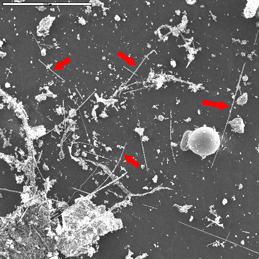
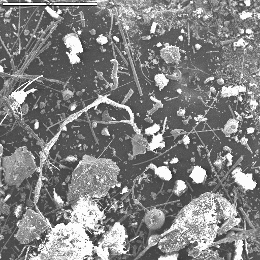
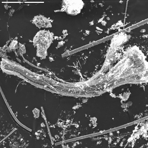
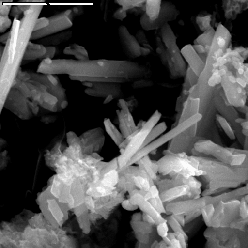
SEM Figure 1 is a scanning electron microscope (SEM) image of a representative portion of sample 22, collected from an area near the World Trade Center. SEM Figure 2 is an image of a representative portion of sample 3 collected near Battery Park. Both images show abundant glass fibers along with particulate debris. SEM Figure 3 is an image of a bundle of chrysotile asbestos from sample 8. The chrysotile was found as a trace constituent in two of the samples analyzed so far by SEM/EDS and in several others analyzed by XRD. SEM Figure 4 is a SEM image of gypsum and/or anhydrite particles consistent with dry wall material. Representative analyses of some of these phases are given in Table SEM-1. Images from other samples can be seen in the Integration of Results section.
Sample 36 was recovered from an indoor location near the Trade Center complex and had not been affected by rain as were the outdoor samples. This sample was thoroughly analyzed for metal-rich particles to better understand the types of materials from which metals might be leached (see leachate studies section). Images of several metal-rich particles can bee seen by clicking here.
Analysis I.D. |
Na2O |
MgO |
Al2O3 |
SiO2 |
SO3 |
K2O |
CaO |
TiO2 |
MnO |
FeO |
Material |
wtc 22 sp 5 |
2 |
11 |
12 |
44 |
trace |
1 |
24 |
1 |
BDL |
4 |
glass fiber |
wtc 22 sp 1 |
trace |
11 |
9 |
47 |
trace |
1 |
32 |
trace |
BDL |
BDL |
glass fiber |
wtc 22 sp 2 |
12 |
2 |
3 |
71 |
trace |
trace |
11 |
trace |
BDL |
trace |
glass fiber |
wtc 3 sp 3 |
trace |
11 |
11 |
43 |
trace |
trace |
32 |
1 |
BDL |
BDL |
glass fiber |
wtc 3 sp 6 |
trace |
11 |
10 |
44 |
trace |
trace |
32 |
trace |
BDL |
1 |
glass fiber |
wtc 8 sp 1 |
BDL |
8 |
9 |
43 |
trace |
trace |
36 |
1 |
BDL |
1 |
glass fiber |
wtc 15 sp 1 |
BDL |
10 |
10 |
42 |
trace |
trace |
36 |
trace |
trace |
BDL |
glass sphere |
wtc 22 sp 4 |
BDL |
12 |
8 |
47 |
trace |
trace |
31 |
BDL |
BDL |
BDL |
glass sphere |
wtc 8 sp 8 |
BDL |
47 |
BDL |
50 |
trace |
BDL |
trace |
BDL |
trace |
2 |
chrysotile |
wtc 14 sp 5 |
BDL |
BDL |
1 |
2 |
53 |
BDL |
43 |
BDL |
BDL |
BDL |
Gypsum/anhydrite |
Data normalized to 100%. BDL=below detection limit. Analytical error is
approximately +/- 5% relative concentration
NEXT Section of Report: Chemical Compositions
Back to document Table of Contents
For more information contact:
Greg Meeker
gmeeker@usgs.gov
| AccessibilityFOIAPrivacyPolicies and Notices | |
| |
|