

Ophidiomycosis (Snake Fungal Disease) Case Definition for Wildlife
Links
- Document: Report (6.3 MB pdf) , HTML , XML
- Larger Work: Case definitions for wildlife diseases
- Download citation as: RIS | Dublin Core
Acknowledgments
The U.S. Geological Survey National Wildlife Health Center and Canadian Wildlife Health Cooperative Case Definition Joint Working Group would like to thank the staff and pathologists in both organizations, as well as our students and colleagues, for the generous contribution of their collective knowledge, expertise, and time creating this case definition.
Introduction
Diagnostic laboratories receive carcasses and samples for diagnostic evaluation and pathogen/toxin detection. The intent of a case definition is to provide scientifically based criteria for determining (1) if an individual carcass has a specific disease and the confidence of that diagnosis; and (2) if a pathogen or toxin is evident in a carcass or sample (for example, swab, tissue sample, skin scraping, blood/serum sample, environmental sample, or other). Using these criteria, cases diagnosed with a specific disease (diagnosing disease) will be classified as “confirmed,” “presumptive,” or “suspected;” and evidence of a pathogen or toxin (detecting pathogen/toxin) will be classified as “exposed” or “present/detected.” Classification is based on a combination of factors: individual, place, time, history, clinical signs, diagnostic observations, and (or) diagnostic test results. Case definitions can bring clarity and consistency to the evaluation process. Their use within and between organizations allows more uniform reporting of diseases and etiologic agents.
Case definitions are proposed for use in wildlife diagnostic laboratories and are not intended to replace regulatory standards provided by Government reporting agencies. Ideally, case definitions would be updated periodically as new information becomes available and new test methods are developed. Refer to the glossary for terminology definitions.
Disease/condition.—Ophidiomycosis (snake fungal disease)
Pathogen/toxin etiologic agent.—Ophidiomyces ophidiicola
Case Definition Criteria
The case definition criteria are a concise summary of the current science regarding the clinical signs, history, gross and microscopic observations, and laboratory test results associated with a specific disease or pathogen. Various combinations of the criteria result in different case classifications representing the degree of certainty of the diagnosis.
Individual, Place, and Time Criteria for Diagnosis and Testing
Individual.—All snake species.
Place.—No restrictions. To date, ophidiomycosis has been found in eastern North America, including the province of Ontario in Canada and the eastern United States and Texas (Baker and others, 2019; Davy and others, 2021).
Time.—No restrictions. In free-ranging snakes, ophidiomycosis seems to be most prevalent in the spring after emergence from brumation (hibernation) (Baker and others, 2019).
Field Criteria for Diagnosis
History and clinical signs.—Clinical signs of ophidiomycosis are highly variable. The most consistent diagnostically compatible signs include scabs or crusty scales, subcutaneous nodules, abnormal molting, white opaque cloudiness of the eyes (not associated with molting), and localized thickening or crusting of the skin. In some cases, there may be skin ulcers, swelling of the face, and nodules in the deeper tissues of the head or body. Ophidiomycosis may cause abnormal feeding leading to emaciation and inappropriate basking behavior, which greatly increases the risk of predation and road mortality, and may expose infected snakes to potentially lethal temperatures (Baker and others, 2019; Davy and others, 2021).
Other.—Not applicable.
Laboratory Criteria for Diagnosis
Gross examination.—Diagnostically compatible postmortem findings include focal to multifocal cutaneous scabbing, crusting, erosion, ulceration, vesicle formation, subcutaneous nodules, or swelling/malformation of the head or body. Dysecdysis or retained spectacles may be present. Affected snakes may be emaciated. Firm nodules (fungal granulomas) may be present in viscera such as a lung or liver (Baker and others, 2019).
Histopathology.—Consistent histopathological findings include epidermal necrosis, particularly within the superficial to mid-epidermis, epidermal erosion or ulceration, heterophilic to lymphoplasmacytic dermatitis, and dermal, pannicular, or muscular granulomas. Ophidiomyces ophidiicola hyphae are visible on periodic acid-Schiff or Grocott methenamine silver stain within areas of necrosis and granulomas, and arthroconidia may be present on the skin surface, but both can be absent within lesions. Hyphae are 2–6 micrometers wide, parallel walled, and septate with acute angle branching. Arthroconidia are rectangular and about 2 by 5 micrometers (Baker and others, 2019).
Diagnostic test(s).—Quantitative polymerase chain reaction (qPCR), polymerase chain reaction (PCR), or fungal culture (Bohuski and others, 2015).
Laboratory Criteria Categorization
Laboratory confirmed.—Positive qPCR and (or) fungal culture for Ophidiomyces ophidiicola; and compatible histopathological lesions, including intralesional fungal elements.
Laboratory supportive.—Based on the following criteria:
-
1. Gross and histologic lesions of ophidiomycosis are present, including characteristic arthroconidia, but O. ophidiicola qPCR or fungal culture is equivocal, negative, or not performed.
-
2. Gross lesions of ophidiomycosis are present and O. ophidiicola qPCR or fungal culture is positive but histopathology is not performed.
-
3. Only scale clip lacking dermis or superficial crust available, but sample contains fungi with morphology consistent with O. ophidiicola and qPCR or fungal culture is positive.
Exposed.—Not applicable.
Present/detected.—Ophidiomyces ophidiicola is detected either by PCR or fungal culture on an individual snake with no gross or histologic lesions compatible with ophidiomycosis; or O. ophidiicola is detected by PCR or fungal culture when no gross lesions are present and histopathology is not performed.
Supplemental Diagnostic Information
Additional diagnostic comments.—Presence of Ophidiomyces ophidiicola can be demonstrated with PCR or fungal culture. Suitable samples for PCR or culture include swabs of the skin, shed skin, scute clips, and (or) biopsies. A confirmed diagnosis of ophidiomycosis can be complicated by the difficulty of morphologically distinguishing O. ophidiicola from other fungi that may cause skin mycoses based on histopathology. In addition, O. ophidiicola may occur on the skin of some snakes in the absence of infection; thus, it is possible that O. ophidiicola could be detected on the skin of some snakes that have mycotic infections caused by other fungi, especially when highly sensitive detection methods such as qPCR are used. Infection with other fungi is more common in captive snakes than in free-ranging snakes because captive snakes are more likely to be infected with other fungi with similar arthroconidia as O. ophidiicola. (Baker and others, 2019; Davy and others, 2021).
Laboratories are encouraged to establish thresholds for qPCR that are likely to reduce the rate of false positive cases of “Laboratory Confirmed” ophidiomycosis based on potential contamination or incidental detection of O. ophidiicola. Refer to Bohuski and others (2015) for more information.
Notifiable/reportable disease.—Not applicable.
Epidemiologic Linkage Criteria for Diagnosis
An epidemiologic linkage can be established by close geographic and temporal proximity (in other words, part of the same mortality event) as one or more confirmed cases of ophidiomycosis or at a site with a recent history of confirmed ophidiomycosis with similar presentation as described in the “Case Definition Criteria” sections.
Case Classification
The sum of the criteria listed in the “Case Definition Criteria” sections (individual, place, time, field, laboratory, and epidemiologic linkage criteria) associated with a particular disease or pathogen/toxin in an individual animal or specimen add up to a case classification (fig. 1; table 1).
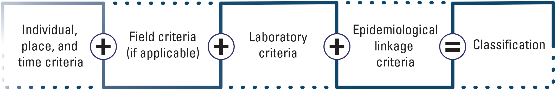
Case definition criteria add up to the case classifications. From Miller and others (2024).
Depending on the confidence in the results, cases of a specific disease will be classified as “confirmed,” “presumptive,” or “suspected;” and evidence of a pathogen or toxin will be classified as “exposed” or “present/detected” (table 1; refer to glossary for definitions). A specific case classification may have more than one pathway to it. Not all classifications may be used for every disease. Compatible epidemiological linkage criteria are required for the “suspected” case classification.
Note.—The field and laboratory criteria in table 1 reflect the typical presentation of snake fungal disease. The exact presentation in an individual animal or specimen may vary from what is presented in table 1 but still conforms with the information presented in the “Field Criteria for Diagnosis” and “Laboratory Criteria for Diagnosis” sections.
Table 1.
Case classification chart for ophidiomycosis (snake fungal disease) and Ophidiomyces ophidiicola.The exposed case classification is not applicable to this case definition.
Quality Assurance Review Schedule
The Canadian Wildlife Health Cooperative and the U.S. Geological Survey National Wildlife Health Center staff plan to review this case definition periodically to incorporate new scientific information and test methods as needed.
Planned date for next review.—June 1, 2025
Review schedule.—June 2025 and then every 3–5 years—or sooner if science about ophidiomycosis (snake fungal disease) changes substantially.
Impact
Applying case definitions in diagnostic, surveillance, and research efforts can help standardize data, making it easier to understand and analyze within and between diagnosticians and laboratories. Laboratories are encouraged to store the case classification assigned to each specimen or sample in their data system so that it can be readily and reliably retrievable.
References Cited
Bohuski, E., Lorch, J., Griffin, K., and Blehert, D., 2015, TaqMan real-time polymerase chain reaction for detection of Ophidiomyces ophidiodiicola, the fungus associated with snake fungal disease: BMC Veterinary Research, v. 11, no. 1, art. 95. [Also available at https://doi.org/10.1186/s12917-015-0407-8.]
Davy, C.M., Shirose, L., Sigler, L., Campbell, D., Dillon, R., Mckenzie, C., Nemeth, N.M., Braithwaite, T., Cai, H., Degazio, T., Dobbie, T., Egan, S., Fotherby, H., Litzgus, J., Manarome, P., Marks, S., Paterson, J.E., Slavic, D., Slavik, E., Urquhart, J., and Jardine, C., 2021, Revisiting ophidiomycosis (snake fungal disease) after a decade of targeted research: Frontiers in Veterinary Science, v. 8, p. 665805. [Also available at https://doi.org/10.3389/fvets.2021.665805.]
Miller, K.J.G., Parmley, E.J., Ballmann, A., Buckner, J., Jones, M., Lankton, J.S., Zimmer, M., and Lankau, E., 2024, [Disease/condition] case definition [template] for wildlife: U.S. Geological Survey Techniques and Methods, book 19, chap. A1, 8 p., https://doi.org/10.3133/tm19A1.
Glossary
additional diagnostic comments
Any additional diagnostic notes pertinent to recording/reporting (for example, requests for strain/serovar/variant reporting, inconclusive/ambiguous results, or “not applicable”).
case classification
The sum of the factors in the “Case Definition Criteria” sections of the case definition including individual (for example, species, age group), place, time, history, clinical signs, diagnostic observations, and (or) diagnostic test results, associated with a particular disease or pathogen/toxin in an individual animal or specimen. Depending on the confidence in the results, cases of a specific disease will be classified as “confirmed,” “presumptive,” or “suspected;” and a pathogen or toxin will be classified as “exposed” or “present/detected.”
case definition
A consistently applied, scientifically based and clearly defined set of field, gross, histopathology, laboratory, or epidemiologic criteria used to classify an individual animal or sample to a specific disease or pathogen/toxin for surveillance or outbreak reporting purposes (based on the combination of the criteria and confidence in the results).
confirmed case
The combination of individual (for example, species, age group), place, time, history, clinical signs, and laboratory criteria for diagnosis with the highest level of certainty for accepted diagnostic testing as stated in the case definition. Example: Cardinal with clinical signs, gross and microscopic lesions compatible with salmonellosis, and positive bacterial culture for Salmonella enterica enterica in the liver.
diagnostic test(s)
Laboratory tests typically used to determine this diagnosis or detect the pathogen/toxin; for example, bacterial culture.
diagnostically compatible
An animal that meets the individual (for example, species, age group), place, time, field, and laboratory criteria for a particular disease as stated in the case definition.
disease
Any disorder of structure or function that produces specific signs or symptoms; disease can be infectious or noninfectious.
disease agent
Any pathogen, toxin, or other known cause of disease.
epidemiologically linked
A case that has temporal, geographic, or other relevant linkages to one or more confirmed cases as described under “Epidemiologic Linkage Criteria for Diagnosis” in the case definition.
exposed
Detection of a toxin in tissues or body fluids at a concentration above acceptable background levels but below the documented lethal threshold level for the species. This may apply to a toxin detected in the absence of documented lethal threshold levels. This category can also include serological evidence of infection in the absence of other information such as organism detection or disease diagnosis.
gross examination
Gross necropsy observations in a carcass or sample that are diagnostically compatible with disease.
histopathology
General microscopic observations in a carcass or sample that are diagnostically compatible with disease.
history and clinical signs
Field observations or changes to behavior in live animals/populations that are diagnostically compatible with disease. Photograph or video evidence may be used when appropriate.
individual
The common age groups, species, or other characteristics that increase disease or pathogen/toxin suspicion.
laboratory confirmed
The strongest degree of assurance in identification of a disease agent of interest and evidence of the associated disease based on one or more accepted laboratory methods. A test or combination of methods that has been scientifically accepted as definitive for a particular disease agent and the associated disease. Example: Positive bacterial isolation for salmonella plus compatible gross and histologic lesions for salmonellosis.
laboratory criteria for diagnosis
The gross, microscopic, molecular, culture, analytical or other laboratory test criteria used to determine the presence of a specific disease agent and evidence of the disease itself. These are categorized based on the validity and performance of the test(s). Categories are “laboratory confirmed,” “laboratory supportive,” “exposed,” and “present/detected.” Where possible, references for the current accepted science for a given disease and pathogen are provided in the case definition. For some select new or emerging diseases the laboratory criteria may be based on the collective expertise of pathologists at the U.S. Geological Survey National Wildlife Health Center and the Canadian Wildlife Disease Cooperative or other institutions.
laboratory supportive
Laboratory results that are less than definitive for a specific disease agent and the associated disease. A test or combination or methods whose results support the diagnosis or a particular disease but are not considered definitive; for example, a screening test. Test result interpretation may be based on the tissue tested (for example, culture of amphibian skin surface versus internal tissue) or postmortem condition of the sample. Example: Gross and histologic lesions compatible with salmonellosis (without laboratory testing).
notifiable/reportable disease
A disease or pathogen that by law must be disclosed to State, Provincial, and (or) Federal agricultural or public health authorities.
other (field criteria)
Additional pertinent comments about presentation (for example, potential for carrier status).
place
Locations and other geographic features that increase disease or pathogen/toxin suspicion.
present/detected
Laboratory detection of a potentially pathogenic agent in the absence of findings diagnostically compatible with the associated disease. Often used when tracking a known or suspected asymptomatic carrier state (for example, Salmonella or duck virus enteritis) or when documenting detection of an agent that is of increased diagnostic or epidemiologic interest, even in the absence of evidence of illness (for example, new or emerging disease or syndrome).
presumptive case
The combination of individual (for example, species, age group), place, time, history, clinical signs and laboratory criteria for diagnosis that has a moderate degree of certainty as stated in the case definition. This uncertainty may be due to the test performed, postmortem decomposition of the carcass affecting observation or interpretation of gross and or histopathologic lesions, inadequate sample for testing due to scavenging or carcass size, inconclusive test results, or lack of a definitive diagnostic test. Enough information is available to conclude the disease is most likely present but not enough information available to conclude the disease is definitively present. Example: Raccoon with compatible histologic lesions for parvovirus without additional laboratory test results.
scope
Indicates what species, when and (or) where this protocol applies; for example, specifics regarding the disease agent, animal class, sex, age group, location, season, antemortem or postmortem sample collection, environmental samples, and so on.
suspected case
This is primarily based on a combination of individual, place, time, minimal or nonspecific field and laboratory information and a geographic and temporal (epidemiologic) connection to a confirmed case. There is not enough information available to meet the threshold in the case definition for a confirmed or presumptive case, but the diagnosis can reasonably be inferred by the close association with confirmed cases of a particular disease in other animals collected from the same general location and time. Example: A specimen with a geographic or temporal link to a confirmed case of a disease that is not tested but was examined and may have nonspecific gross or histopathologic findings that are compatible with that disease.
time
The season(s), months, or other temporal factors that increase disease or pathogen/toxin suspicion.
- wildlife
Free ranging vertebrate species (mammals, birds, reptiles, amphibians, and fish).
For more information about this publication, contact:
Director, USGS National Wildlife Health Center
6006 Schroeder Road
Madison, WI 53711
For additional information, visit: https://www.usgs.gov/centers/nwhc
Publishing support provided by the Rolla Publishing Service Center
Disclaimers
Any use of trade, firm, or product names is for descriptive purposes only and does not imply endorsement by the U.S. Government.
Although this information product, for the most part, is in the public domain, it also may contain copyrighted materials as noted in the text. Permission to reproduce copyrighted items must be secured from the copyright owner.
Suggested Citation
Lankton, J.S., Stevens, B., Shirose, L., and Davy, C., 2024, Ophidiomycosis (snake fungal disease) case definition for wildlife: U.S. Geological Survey Techniques and Methods, book 19, chap. F1, 8 p., https://doi.org/10.3133/tm19F1.
ISSN: 2328-7055 (online)
| Publication type | Report |
|---|---|
| Publication Subtype | USGS Numbered Series |
| Title | Ophidiomycosis (snake fungal disease) case definition for wildlife |
| Series title | Techniques and Methods |
| Series number | 19-F1 |
| DOI | 10.3133/tm19F1 |
| Publication Date | March 15, 2024 |
| Year Published | 2024 |
| Language | English |
| Publisher | U.S. Geological Survey |
| Publisher location | Reston, VA |
| Contributing office(s) | National Wildlife Health Center |
| Description | v, 8 p. |
| Online Only (Y/N) | Y |
| Additional Online Files (Y/N) | N |